
Progressive retinal atrophy (PRA)
From Wikipedia, the free encyclopedia
Progressive retinal atrophy (PRA) is
a group of genetic diseases seen in certain breeds of dogs and, more rarely, cats. Similar to retinitis pigmentosa in humans,it is characterized by the bilateral degeneration of the retina,
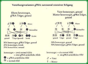
causing progressive vision loss culminating in blindness. The condition in nearly all breeds is inherited as an autosomal recessive trait.
Types of PRA:
In general, PRAs are characterised by initial loss of rod photoreceptor cell function followed by that of the cones and for this reason night blindness is the first significant clinical sign for most dogs affected with PRA. As other retinal disorders, PRA can be divided into either dysplastic disease, where the cells develop abnormally, and degenerative, where the cells develop normally but then degenerate during the dog's lifetime.
Generalized PRA is the most common type and causes atrophy of all the neural retinal structures. Central progressive retinal atrophy (CPRA) is a different disease from PRA involving the retinal pigment epithelium (RPE), and is also known as retinal pigment epithelial dystrophy (RPED).
Rod-cone dysplasia type 2
Collie - Rod cell response is nearly absent. Night blindness by six weeks old, blind by one to two years old.
Central progressive retinal atrophy (CPRA)
CPRA is also known as retinal pigment epithelial dystrophy (RPED). The cause of this condition is the loss of the retinal pigment epithelium's ability to effectively process the photoreceptor outer segment (POS) and subsequent accumulation of POS material in the RPE and loss of function. The loss of function of the RPE leads to photoreceptor degeneration. Vitamin E deficiency may play a role in the development of CPRA. It is characterized by accumulation of pigment spots in the retina surrounded by retinal atrophy and a mottled appearance of the pigmented nontapetal fundus. The pigmented spots eventually coalesce and fade as the atrophy of the retina increases. It is an inherited condition (in the Labrador Retriever it is inherited as an autosomal dominant trait with variable penetrance). CPRA occurs in older dogs. Peripheral vision is retained for a long time. Vision is better in low light and better for moving or distant objects. Not all affected dogs go blind. Secondary cataracts are common.
Diagnosis
Progressive vision loss in any dog in the absence of canine glaucoma or cataracts can be an indication of PRA. It usually starts with decreased vision at night, or nyctalopia. Other symptoms include dilated pupils and decreased pupillary light reflex. Fundoscopy to examine the retina will show shrinking of the blood vessels, decreased pigmentation of the nontapetal fundus, increased reflection from the tapetum due to thinning of the retina, and later in the disease a darkened, atrophied optic disc. Secondary cataract formation in the posterior portion of the lens can occur late in the disease. In these cases diagnosis of PRA may require electroretinography (ERG). For many breeds there are specific genetic tests of blood or buccal mucosa for PRA.
Absent a genetic test, animals of breeds susceptible to PRA can be cleared of the disease only by the passage of time—that is, by living past the age at which PRA symptoms are typically apparent in their breed. Breeds in which the PRA gene is recessive may still be carriers of the gene and pass it on to their offspring, however, even if they lack symptoms, and it is also possible for onset of the disease to be later than expected, making this an imperfect test at best.
OUR COLLIES ARE TESTED BY ECVO & DNA. Unsere Collies sind klinisch und genetisch getestet und PRA frei!